Dados Clínicos
Sexo feminino, 1 ano e 7 meses.
HDA: Histórico de náuseas e vômitos há 7 dias. Episódios recorrentes de ITU, fez uso de
diversos antimicrobianos (bactrim, cefadroxila, ceftriaxona e cefuroxima).
HPP e HFS: Parto normal, a termo. Sem intercorrências gestacionais conhecidas.
Exame físico:
Anisocoria(D>E), Sem fotorreação à direita. Sem rigidez de nuca. Paralisia facial central, nistagmo horizontal , bem como hiperreflexia bilateral em membros inferiores.
Exames complementares
- Laboratório: Leucocitose (18800), sem desvio. PCR 40 mg/L
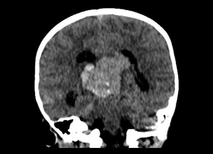
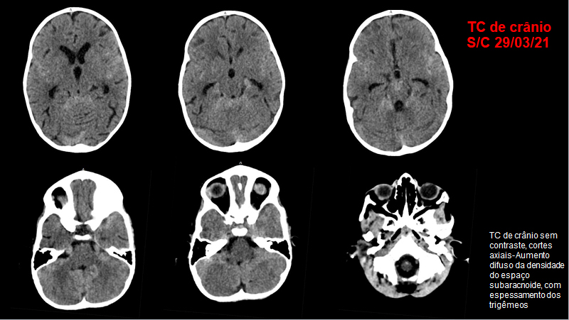
Solicitada TC de crânio (29/03)
Rangel de Sousa Costa
Fellow de Neurorradiologia do Instituto Estadual do Cérebro (IEC)
Paulo Niemeyer (R5)
Orientação: Gabriela de Almeida Lima e Nina Ventura.

Legendas
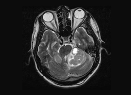
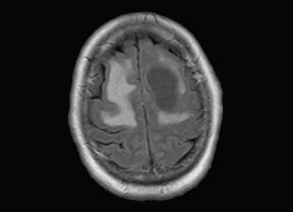
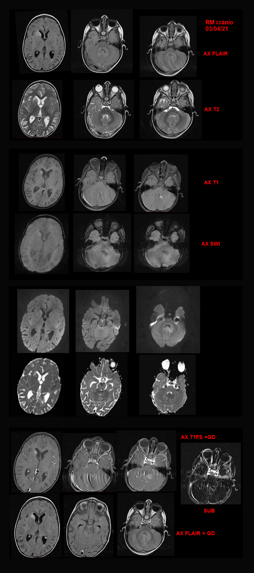
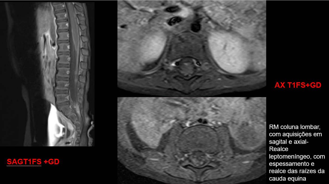
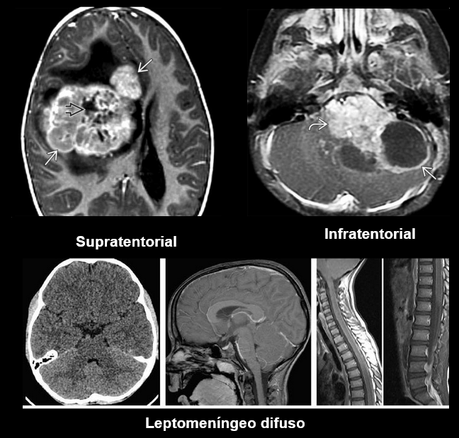
Lesões leptomeníngeas difusas, com impregnação pelo contraste e restrição à difusão. Nota-se ainda lesão mal definida no hemisfério cerebelar esquerdo, com foco de sangramento de permeio.
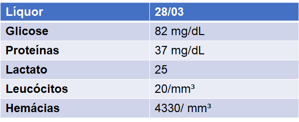
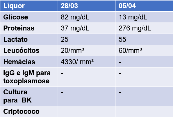
Realizada nova punção liquórica
Conclusão
Liquor com células neoplásicas de histogênese incerta.
Paciente submetida a biópsia da lesão superficial no cerebelo, que foi inconclusiva.
O diagnóstico foi fechado no estudo de autópsia: Tumor teratoide rabdoide atípico, com apresentação predominantemente leptomeníngea.
Tumor Teratoide Rabdoide Atípico
- Tumor embrionário, descrito inicialmente em 1987;
- Geralmente acomete crianças até 2 anos;
- Agressivo, frequentemente letal (sobrevivência em 3 anos de 5% quando infratentorial);
- Mais comum em meninas;
- Intraxial (94%);
- Tanto supra quanto infratentorial;
- Infratentorial: mais comum de linha média, ângulo ponto cerebelar, cerebelo/tronco encefálico.
Supratentorial: geralmente hemisférico;
Achados de imagem:
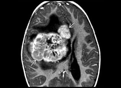
- Geralmente se apresenta como massa > 3cm;
- TC: Massa hiperdensa; comumente com cistos e hemorragia; Pode apresentar calcificações;
- RM: Geralmente isointenso ao parênquima cerebral; focos hemorrágicos (blooming no T2*). Marcada restrição à difusão.
- Realce heterogêneo;
- Disseminação meníngea comum (15-20% no diagnóstico inicial)
Achados de imagem
- Bhimani AD et al: Differentiation of outcomes by treatment regimen and histology in CNS primary embryonal tumors. World Neurosurg. ePub, 2020
- Blessing MM et al: Embryonal tumors of the central nervous system: an update. Surg Pathol Clin. 13(2):235-47, 2020
- Cacciotti C et al: Advances in the molecular classification of pediatric brain tumors: a guide to the galaxy. J Pathol. ePub, 2020
- Li G et al: Screening for genes and subnetworks associated with atypical teratoid/rhabdoid tumors using bioinformatics analysis. Int J Neurosci. 1-8, 2020
- Pan X et al: Identification of hub genes in atypical teratoid/rhabdoid tumor by bioinformatics analyses. J Mol Neurosci. ePub, 2020
- Kristensen BW et al: Molecular pathology of tumors of the central nervous system. Ann Oncol. 30(1):1265-78, 2019
- Nesvick CL et al: Case-based review: atypical teratoid/rhabdoid tumor. Neurooncol Pract. 6(3):163-78, 2019
- Oh CC, Orr BA, Bernardi B, et al. Atypical teratoid/rhabdoid tumor (ATRT) arising from the 3rd cranial nerve in infants: a clinical- radiological entity? J Neurooncol 2015;124(2):175–183.
- El-Nabbout B, Shbarou R, Glasier CM, Saad AG. Primary diffuse cerebral leptomeningeal atypical teratoid rhabdoid tumor:report of the first case. J Neurooncol 2010;98(3):431–434.